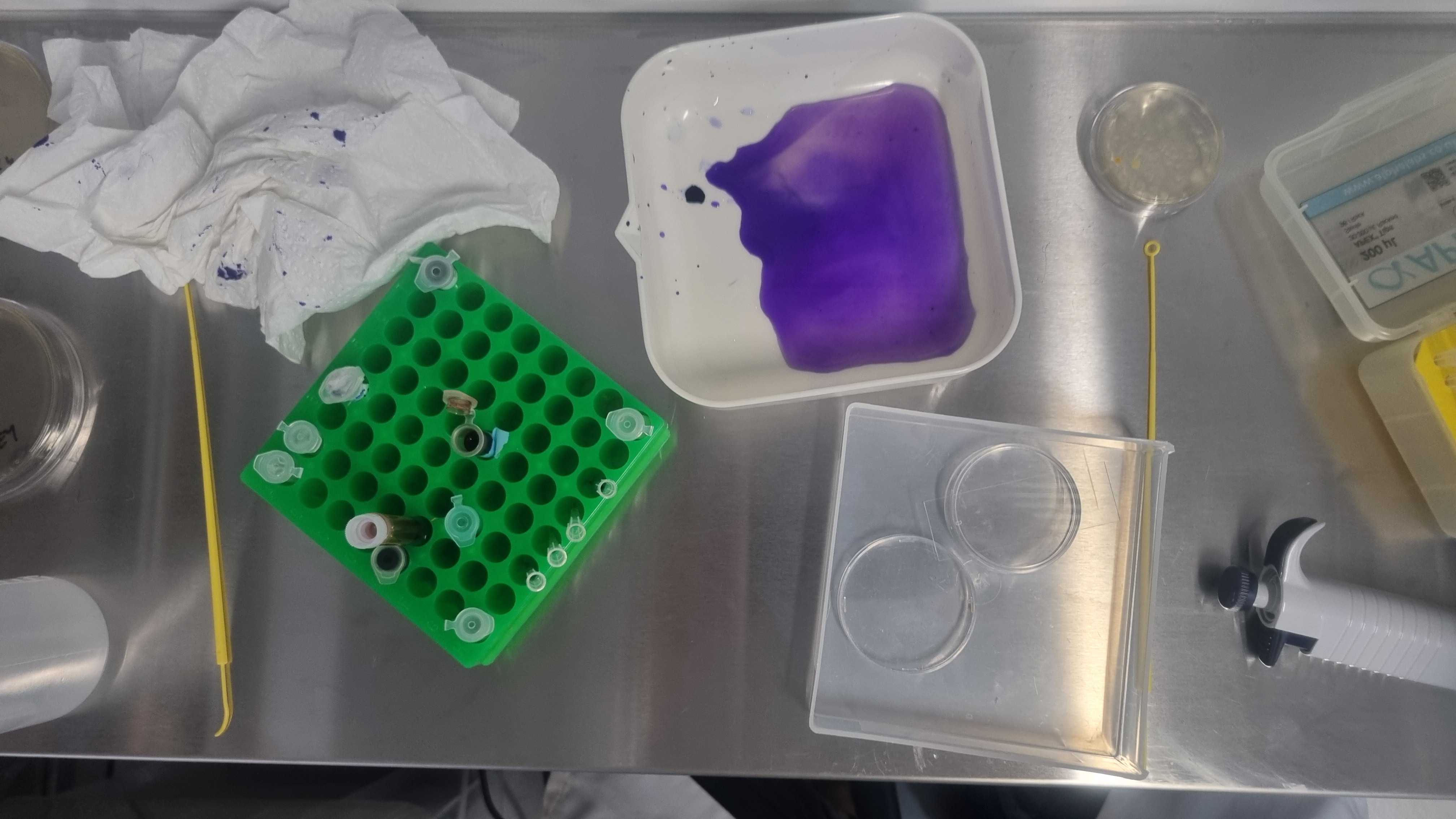
Gram-Staining
make your bacteria purple

A beautiful technique for seeing, indentifying and working with bacteria from samples.
Named after Hans Christian Gram who invented the procedure in 1884.
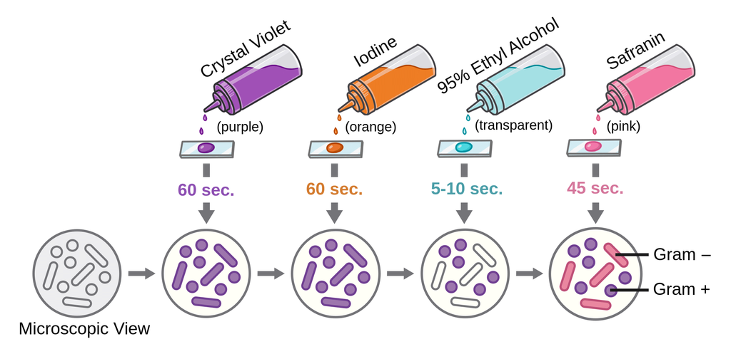
The procedure classifies bacteria into two large groups: gram-positive or gram-negative based on the physical properties of their cell walls, stainign them either purple (positive) or pink (negative). Gram-positive bacteria respond better to wall-targeting antibiotis due to their lack of outer membrane (which is also why they take the stain so well).
Once you can see bacteria you can infer:
- Abundance (how much)
- Evenness (how is it distributed)
- Moprhology (what does it look like)
- Taxonomy (sometimes) (who is it?)
You can also use it to differntiate between bacteria and fungi .

Needed equipment:
- sample
- microscope plate
- innoculation loop
- bunsen burner
- pipette
- chemicals:
- Crystal Violet
- Iodine
- Alcohol
- Safronin
- Water
Steps:
- Assemble all gear and get a plate
- Sterilise loop
- Swab sample
- Transfer sample to plate (if using water run loop through 20UL of water on plate)
- Fix to plate by passing it over the burner three times
- Apply chemical and rinse with water between each one (see picture) :
| Crystal Violet | Iodine | Alcohol | Safronin |
| 1 min | 1 min | 10 secs | 1 min |
Allow to dry then observe under microscope.